


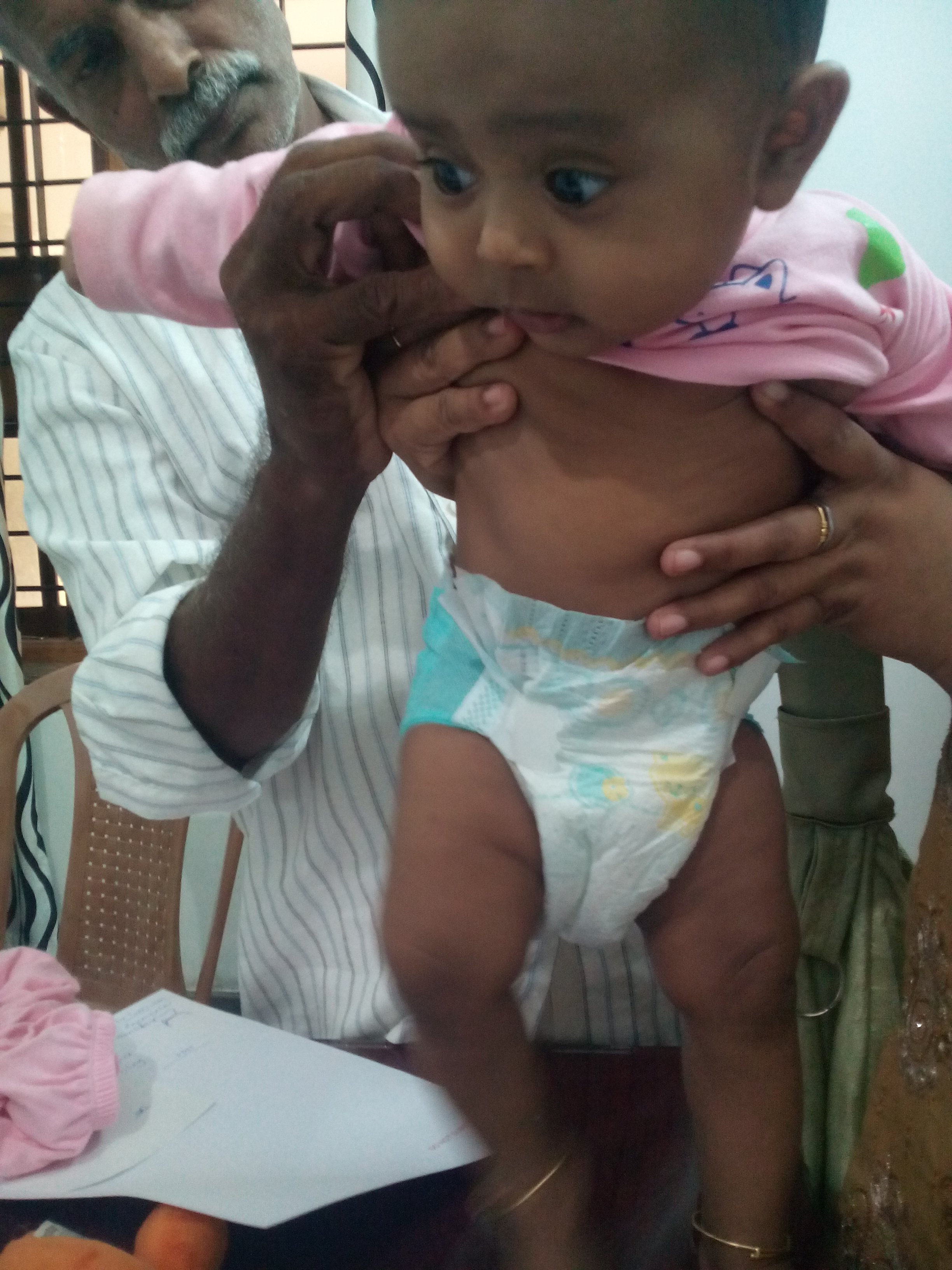
- 5 month girl is brought with h/o rt foot is placed as tip toe whenever she bounces on feet
- NO H/O TRAUMA/LOCAL INJURY
- birth h/o FTND
- O/e rt foot equinus, no contracture,no other bony deformity
- leg length difference 1.5 cm, tone reflexes normal,spine normal
- hip abduction limited on rt side
- extra skin fold noted by parents
suggested
pelvis ap
frog leg lateral
dx:ddh untreated till 5 months with shortening
CRITIQUE: identify a treatable cause as early as possible -DDH
PARENT HAS ONLY ONE QUESTION-WILL INTERVENTION LEAVE HER WITHOUT PROBLEM?
- Management
Infants (newborn to 2 weeks old) who have a positive finding on an Ortolani or Barlow examination should be referred urgently (within 1–2 weeks) to an orthopedist comfortable with the management of DDH. Infants age 2 weeks who have equivocal findings may undergo ultrasonography or be evaluated by an orthopedist to confirm whether or not a dislocated hip is present. - Initial management of DDH consists of the use of a hip abduction brace, usually a Pavlik harness in infants younger than age 6 months. Brace management protocols vary, but typically involve a period of full-time harness wear followed by nighttime wear. Frequent follow-up visits are necessary to adjust the harness and prevent complications resulting from a harness that is too small, specifically neuropathies and AVN
- Primary care physicians often play a role in initial identification of neuropathies, especially for families living far from the treating orthopedist. Parents are advised to look for symmetric, spontaneous movement of all extremities whenever the harness is removed. If abnormalities are seen, the harness should be left off and the child should be evaluated by the orthopedist shortly thereafter. Pavlik harness treatment is most successful when initiated within the first 2 months after birth.
- Early treatment with the Pavlik harness results in reduced and stable hips in more than 95% of all cases of DDH, although complete dislocations are more likely to fail treatment. Because hips that remain dislocated invariably progress to poor outcomes as early as young adulthood, hips that fail to reduce and stabilize with Pavlik harness treatment require reduction under anesthesia and hip spica casting. Often, onlyminimal surgical intervention is needed, but more extensive procedures may be necessary to correct any underlying abnormalities that prevent reduction
- Children who present at a later age should be referred within a few weeks to an orthopedic surgeon. Radiographic dysplasia may be addressed with rigid hip abduction bracing in children older than age 6 months, butlate-presenting dislocations require surgical reduction,just as with teratologic dislocations.Patients who have unresolved radiographic dysplasiaor residual dysplasia, after operative or nonoperativetreatment, benefit from additional surgical procedures to improve the relationship between the acetabulumand femoral head before skeletal maturity. umerous procedures may be used, depending on the age of the patientand the configuration of the acetabulum and femoral head.
- Prognosis
Early identification and treatment of unstable hips with a Pavlik harness generates the best outcomes. The complications of Pavlik harness treatment, AVN and nerve palsies, are rare and are best prevented with close monitoring during treatment. Approximately 1% of patients have residual dysplasia requiring surgery during the first 5 years after birth, (13) and, despite normal radiographic findings at age 4 to 5 years, 10% to 15% may demonstrate dysplasia on radiographs obtained during adolescence. As a result, patients should be followed through skeletal maturity with periodic radiographic surveillance, usually by the treating orthopedist. - Patients in whom Pavlik treatment fails, or who present later in age, and who undergo closed or open reduction of their hips frequently require additional surgery before skeletal maturity, usually during the first 4 to 5 years after birth. The rate of development of osteoarthritis in patients treated for DDH is unknown. Those who have residual dysplasia certainly are at higher risk, but later osteoarthritis is a consideration even in those who have normalappearing hips at skeletalmaturity.Osteoarthritis in patients who have a history of DDH is a leading cause of joint replacement in adults, occurring as young as the third decade after birth.
-
Summary
- Based on strong research evidence, the primary risk factors for developmental dysplasia of the hip (DDH)include female gender, family history, and breech presentation
- Based on current standards of care and published guidelines, all children should receive routine clinical evaluation of their hips at each scheduled health supervision visit.
- Based primarily on consensus due to the lack of relevant clinical studies, children who have equivocal findings or increased risk factors for DDH (and normal examination findings) should undergo imaging with ultrasonography at age 3 to 4 weeks, or plain radiographs at 4 to 5 months if reliable ultrasonography is not available
- Based on strong research evidence, infants who manifest adventitial hip clicks do not require further imaging or referral to an orthopedic surgeon.
- Based on consensus due to the lack of relevant clinical studies, children who have unstable hips on clinical examination should be referred for treatment by an orthopedist.
- Based on consensus due to the lack of relevant clinical studies, children who have abnormal findings on radiographic evaluation, either on ultrasonography or plain radiographs, should be referred to an orthopedist for evaluation and determination of appropriate management.
 Superintendent, ICH.